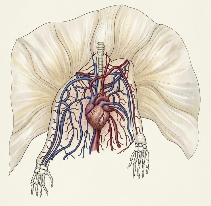
A simplified anatomical visual of the central human circulatory system. Photo credit: Original hand drawing by Shamil Issayev, digitally colored and processed using Vizcom.
By Shamil Issayev ‘28
Approximately 108,000 daily heart contractions, each pumping 70 milliliters or 5.25 liters of blood per minute, traveling through the internal blood vessels, which if were to be separated and connected together, would feature at a confounding 96,500 kilometers or 60,000 miles. This vascular strand is enough to circle the Earth more than twice, and it is known as the human circulatory system (Anatomy and Physiology 2e).
Before touching the idea of the circulatory system as a whole, it is necessary to understand what causes the internal fluids to circulate in the first place. One of the most important organs of the human body and the primary pump in cardiac mechanics is the heart. The heart’s shape and size, specific location, external and internal structures, and functions are what makes it a perfect catalyst for lifelong impulses.
The organ is shaped similar to a pinecone, typically the approximate size of an adult human fist. As identified in Anatomy and Physiology 2e, it is 12 centimeters (5 in) in length, 8 centimeters (3.5 in) in width, and 6 centimeters (2.5 in) in thickness. These dimensions are crucial to hold a stroke volume of roughly 70 milliliters of blood per beat or 5 liters of blood per minute in its ventricles. This way it prevents cellular suffocation and maintains chemical homeostasis. During cardiovascular exercise, when the working muscles demand up to 10 times more oxygen, a healthy heart can increase its pumping rate to 20-25 liters per minute.
It is located within the chest cavity between the lungs in the space known as mediastinum. The back of the heart lies near the spine, while its front sits just behind the rib cage. The top of the heart or the base is leveled with the third rib cartilage, attaching the body’s major vessels to itself. The bottom tip of the heart, the apex, is positioned to the left of the breastbone (sternum) between the fourth and fifth ribs near their connection with the rib cartilage.
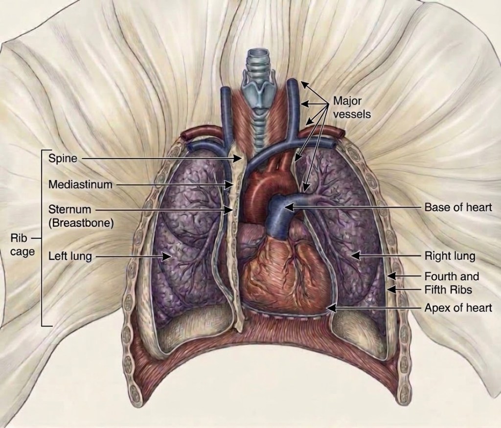
An anatomical diagram of the human heart’s location. Photo credit: Using the idea from Anatomy and Physiology 2e, hand-drawn, digitally colored, and processed using Vizcom by Shamil Issayev.
Externally, the heart is separated from other mediastinal structures by a tough membrane known as the pericardium. The term directly translates as “around the heart” and consists of two distinct sublayers: the durable outer fibrous pericardium and the inner serous pericardium. The fibrous pericardium is made of tough, dense connective tissue that protects the organ and stabilizes its position in the thorax. The serous pericardium consists of its own two layers: the parietal pericardium, which is fused to the fibrous pericardium, and an inner visceral pericardium, or epicardium, which is fused to the heart and is part of the heart wall.
In most organs within the body, visceral serous membranes like the epicardium are typically microscopic; in the heart, however, it is composed of a simple squamous macroscopic layer called a mesothelium, reinforced with loose, irregular connective tissue. These structures establish the organ’s own secure space called the pericardial cavity filled with lubricating fluid.
In the case of a condition called cardiac tamponade or pericardial tamponade, with each contraction of the heart, more fluid—in most instances, blood—accumulates within the pericardial cavity. This puts pressure on the heart and prevents full relaxation to fill the pump for the next contraction. In hypothyroidism, the fluid builds up slowly, and the pericardial cavity may be able to expand gradually to accommodate this extra volume. Other common causes of unnatural pressure include myocardial rupture, pericarditis, cancer, or even cardiac surgery.
The middle and thickest layer of the heart wall is myocardium, mostly composed of cardiac muscle cells. It is structured upon collagenous fibers and the blood vessels that supply the layer and the nerve fibers that help regulate the heart. The pumping of the blood through the heart into major arteries is triggered by the contraction of the myocardium. This muscle pattern is lean and complex, as the muscle cells spiral around the chambers of the heart. The innermost layer of the heart wall is the endocardium, which joins to the myocardium with a thin layer of connective tissue. It forms a smooth, continuous outer line inside where the blood circulates and covers the heart valves without causing friction.
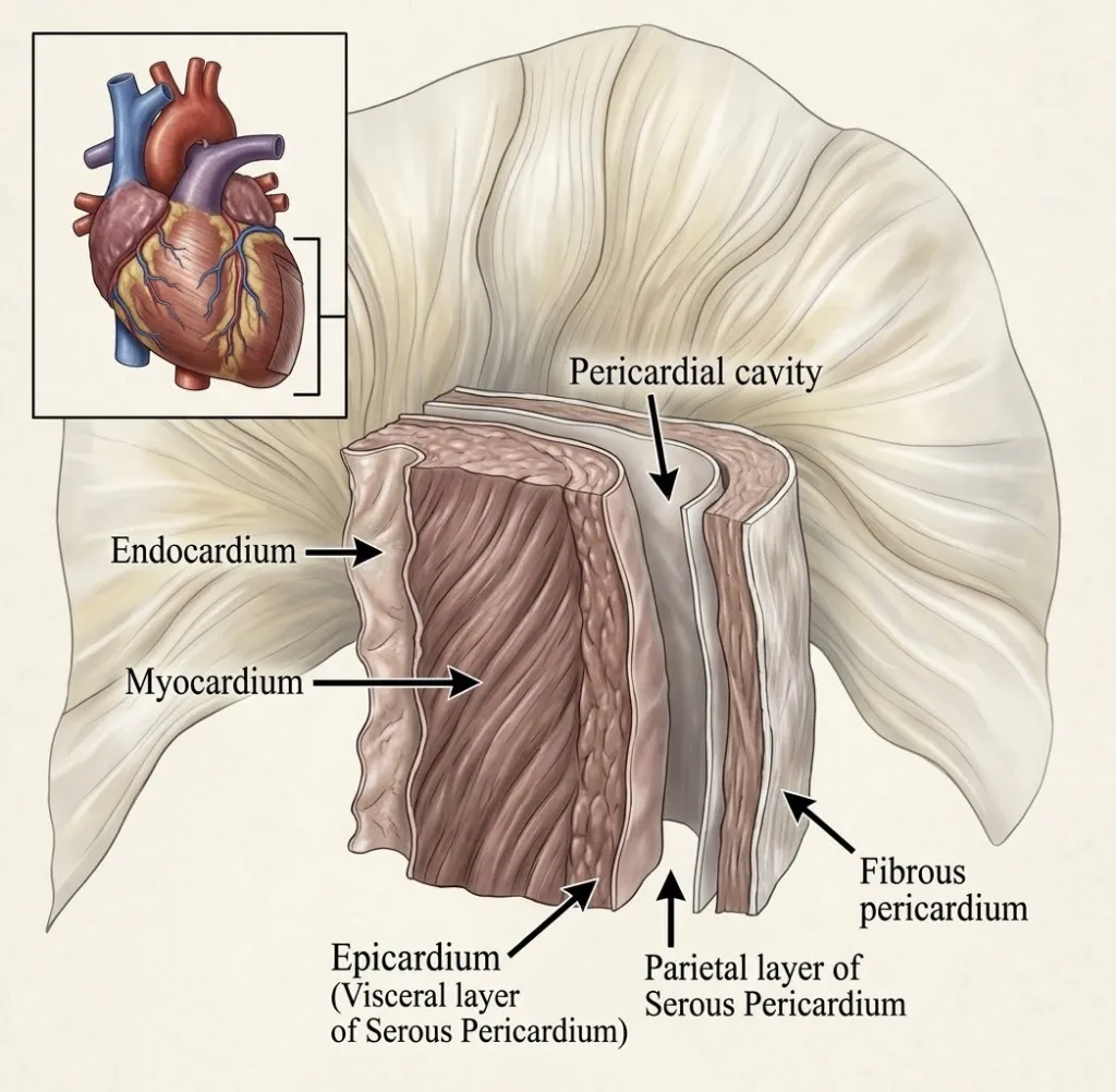
An anatomical diagram of the pericardial membranes and layers of the human heart wall. Photo credit: Using the idea from Anatomy and Physiology 2e, hand-drawn, digitally colored, and processed using Vizcom by Shamil Issayev.
Internally, the heart is partitioned into chambers called atria and ventricles by a muscular wall called the septum, which is the physical extension of the myocardium lined with endocardium. As detailed in Anatomy and Physiology 2e, the interatrial septum divides the receiving atria and bears the fossa ovalis—a remnant of the fetal foramen ovale, which once allowed blood to bypass the lungs.
Within seconds after birth, a flap of tissue known as the septum primum permanently seals this opening to establish postpartum circulation. Between the two ventricles, below, is a second septum known as the interventricular septum, which is normally intact after its formation during fetal development. It is substantially thicker than the interatrial septum because the ventricles generate a significantly greater pressure when they contract.
The septum between the atria and ventricles, which is reinforced with dense connecting tissue called the cardiac skeleton, is known as the atrioventricular septum. It is marked by the presence of four openings that allow blood from the atria into the ventricles and from the ventricles into the pulmonary trunk and aorta—the system’s largest vessel. The valves between the atria and ventricles are generally known as atrioventricular valves, and the valves at the openings that lead to the pulmonary trunk and aorta are known as semilunar valves.
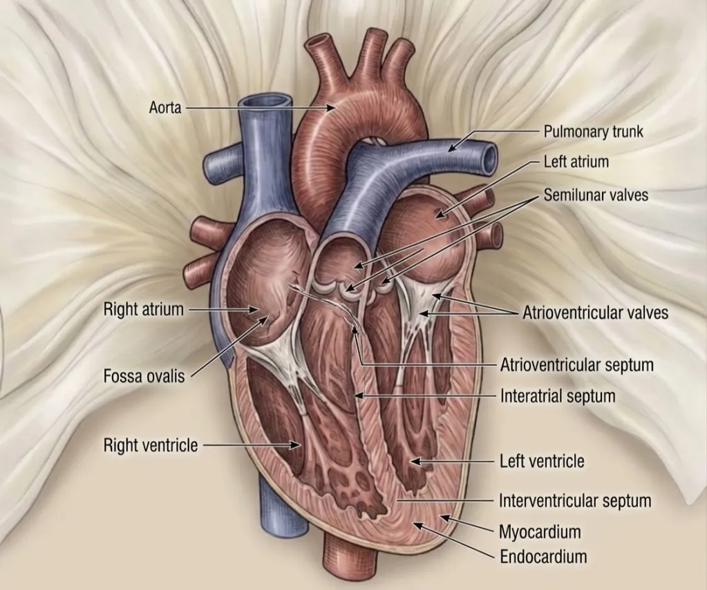
An anatomical diagram of the four chambers and one-way valves of the internal human heart. Photo credit: Using the idea from Anatomy and Physiology 2e, hand-drawn, digitally colored, and processed using Vizcom by Shamil Issayev.
In addition to the heart, the circulatory system is composed of various blood vessels, each possessing their own characteristic and function. There are a total of four general vessel types of macroscopic and microscopic sizes throughout the body. An example of one of the larger types is an artery, which plays a major role in nourishing organs with blood and nutrients. As outlined in “Anatomy, Blood Vessels,” arteries are under constant high pressure, and to accommodate this stress, they have an abundance of elastic tissue and rough muscle. The possession of elastin in such vessels allows them to increase in size and alter their diameter. When arteries reach a particular organ, they undergo further division into smaller vessels. It is estimated that about 10% to 15% of the total blood volume is contained in the arterial system, which features a system of high systemic pressure and low volume.
Consequently, there are two main subtypes of arteries found in the body: the elastic arteries and the muscular arteries. Elastic arteries are the vessels nearest the heart, like the aorta and pulmonary arteries, and contain more elastic tissue in the tunica media than muscular arteries. This feature of the elastic arteries allows them to maintain a relatively constant pressure gradient despite the constant pumping action of the heart. Muscular arteries, on the other hand, possess a limited ability to expand due to a decreased quantity of elastic fibers, which aligns with the already lowered blood pressure as the system reaches these distant subtypes.
In addition to elastic and muscular arteries, a different category of arteries exists—the arterioles. These are the smallest 8–60-micrometer-type of arteries, having their diameter and shape being influenced by the nervous system. They respond to the tissue’s need for nutrients and oxygen, playing an important role in the systemic vascular resistance because of the lack of significant elastic tissue in the walls.
Branching directly from terminal arterioles are capillaries, which are microscopic exchange vessels measuring 5 to 10 micrometers in diameter—the width just enough to allow red blood cells to pass through. As detailed in Anatomy and Psychology 2e, they are composed of a single layer of endothelial cells resting on a basal lamina representing only the tunica intima and lacking both a tunica media and tunica externa, making them the only vessel across the circulatory system where gas, nutrient, and waste exchange occurs through diffusion.
Capillaries are characterized into three distinct structural subtypes based on their endothelial permeability, similar to arteries. An example is a continuous capillary, featuring uninterrupted linings bound by tight junctions in skeletal muscle, skin, and the brain’s blood-brain barriers.
Another subtype is fenestrated capillaries, which possess small membrane-covered pores, or “windows” (fenestrae), allowing rapid fluid and solute filtration in the kidneys, endocrine glands, and small intestine. The last of the subtypes is sinusoidal capillaries, which feature large intercellular gaps, incomplete basal laminae, and wide lumens to permit the passage of large proteins and intact blood cells found in the liver, bone marrow, and spleen.
From capillaries blood enters venules, which are small microvascular conduits that mark the beginning of the venous return system. These types range in diameter from 8 to 100 micrometers and can join to form a vein. Venules are characterized into two primary structural subtypes: postcapillary venules and muscular venules.
The most known of the vessels are veins, which are engineered to channel blood back toward the heart. They are structurally categorized into small-to-medium subtypes such as radial or tibial veins and large veins such as the venae cavae and pulmonary veins. Since these vessels are at low pressure, larger veins are commonly found with essential valves to maintain the unidirectional flow of blood toward the heart. This prevents backflow towards the other vessels caused by the gravitational force.
Lastly, in addition to the four general types, a metarteriole is a blood vessel that is structurally similar to both an arteriole and a capillary. The smooth muscle of the tunica media of the metarterioles is not continuous as in other types, forming muscular rings called sphincters prior to the entrance to the capillaries. Each of these types stems from a terminal arteriole and branches to supply blood to a capillary bed.
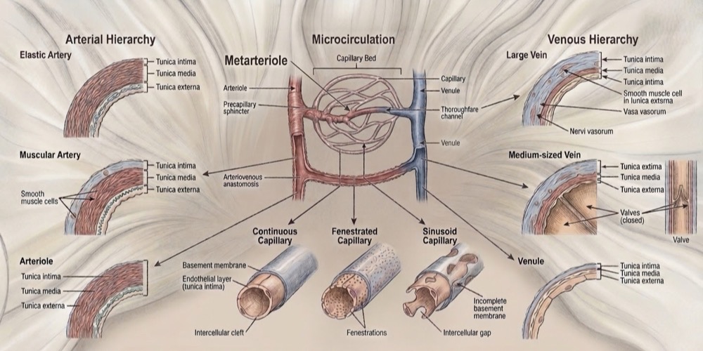
An anatomical diagram of the human blood vessel types. Photo credit: Using the idea from Anatomy and Physiology 2e, hand-drawn, digitally colored, and processed using Vizcom by Shamil Issayev.
Blood circulation is managed through a continuous dual-circuit pathway: the pulmonary circuit and the systemic circuit. According to “How the Heart Works,” in the pulmonary circuit, deoxygenated blood returns through the venae cavae into the right atrium, passes through the tricuspid valve into the right ventricle, and is released through the pulmonary valve into the pulmonary trunk. The flow then changes its vector towards the lungs, where it enters the smooth alveolar capillaries under low pressure of about 25 mmHg to exchange carbon dioxide for oxygen before returning the pulmonary veins to the left atrium.
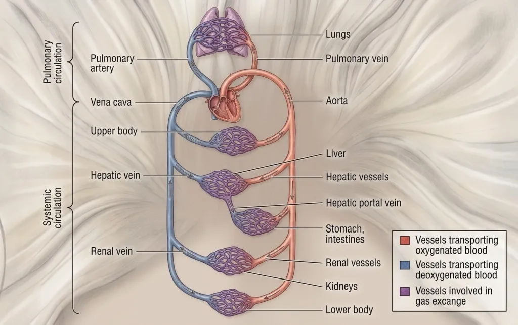
An anatomical diagram of the human vascular dual-circuit pathway. Photo credit: Using the idea from Anatomy and Physiology 2e, hand-drawn, digitally colored, and processed using Vizcom by Shamil Issayev.
The heart depends on specific electrical and mechanical physics in order to manage this complex loop. As outlined in clinical reviews of “Physiology, Cardiac Cycle,” the cardiac cycle originates from the sinoatrial (SA) node, which is the heart’s natural pacemaker located in the upper wall of the right atrium. The sinoatrial node fires spontaneously at 60 to 100 beats per minute, from which the electrical impulse then travels to the atrioventricular (AV) node. The atrioventricular node then creates a crucial delay of approximately 0.1 seconds to give the atria time to contract fully before the ventricles squeeze.
This creates a dynamic four-phase mechanical cycle lasting 0.7 seconds during which the isovolumetric contractions snap the AV valves shut. Following that, rapid ventricular forces open the semilunar valves to push out a 70 milliliter stroke volume, with isovolumetric relaxation snapping the aortic and pulmonary valves shut and the passive ventricular filling finishing with an “atrial” that fills the ventricles to a 120 milliliter end-diastolic volume.
The circulation of blood through these vessels is maintained by strict physiological physics. According to Anatomy and Physiology 2e, blood flow is directly proportional to the pressure gradient and inversely proportional to vascular resistance dictated by Poiseuille’s Law. The law reveals that vessel radius is the single most powerful control point in the entire system.
The resistance is inversely proportional to the radius raised to the fourth power; having an arteriole’s radius increases resistance sixteen times and prevents flow down of a fraction of its original volume. This fluid dynamic was used to derive the clinical metric of mean arterial pressure, where a typical unit of 120/80 mmHg yields a mean pressure of approximately 93.3 mmHg.
Maintaining this vascular equilibrium has grown to a critical global importance. According to standards from “Cardiovascular Basics and Circulation,” a normal blood pressure is below 120/80 mmHg, while a pressure of 130/80 mmHg falls under hypertension thresholds. On a larger scale, statistics from “Global Health Estimates: Leading Causes of Death and Disability” indicate that cardiovascular diseases are the absolute leading cause of death worldwide, estimating 17.9 million lives each year. This is roughly 32% of all global mortality, with over 85% of those deaths stemming directly from heart attacks and strokes linked to vascular failure.
Maintaining this vascular equilibrium is a matter of critical global importance. A normal blood pressure is below 120/80 mmHg, while readings at or above 130/80 mmHg cross into hypertension thresholds. On a macro scale, statistics from “Global Health Estimates: Leading Causes of Death and Disability” indicate that cardiovascular diseases are the absolute leading cause of death worldwide, estimating 17.9 millions lives each year. This is roughly 32% of all global mortality, with over 85% of those deaths stemming directly from heart attacks and strokes linked to vascular failure.
To maintain a healthy circulatory state, it is important to engage in regular physical activities, specifically aerobic exercise, which strengthens the myocardium and enhances the efficiency of blood pumping. Focusing on a heart-healthy eating pattern consisting of nutrient-dense meals rich in fruits, vegetables, fiber-rich whole grains, and lean proteins, while limiting sodium, added sugars, and saturated fats, is the diet recommended by “How the Heart Works.” It is especially important for the current generations to prioritize rest and stress management, which supports overall autonomic balance and long-term vascular health.